
Mammography Clinic
Early Detection Saves Lives
Mammography is a specialised X-ray examination of the breasts designed to detect breast cancer and other breast abnormalities at the earliest possible stage—often before any symptoms are noticeable. Regular screening plays a vital role in improving treatment outcomes and supporting long-term breast health.
At Tshirulu Radiology, we provide high-quality digital mammography in a comfortable, private, and compassionate environment. Our experienced team is committed to ensuring that every patient receives professional care with dignity and respect.
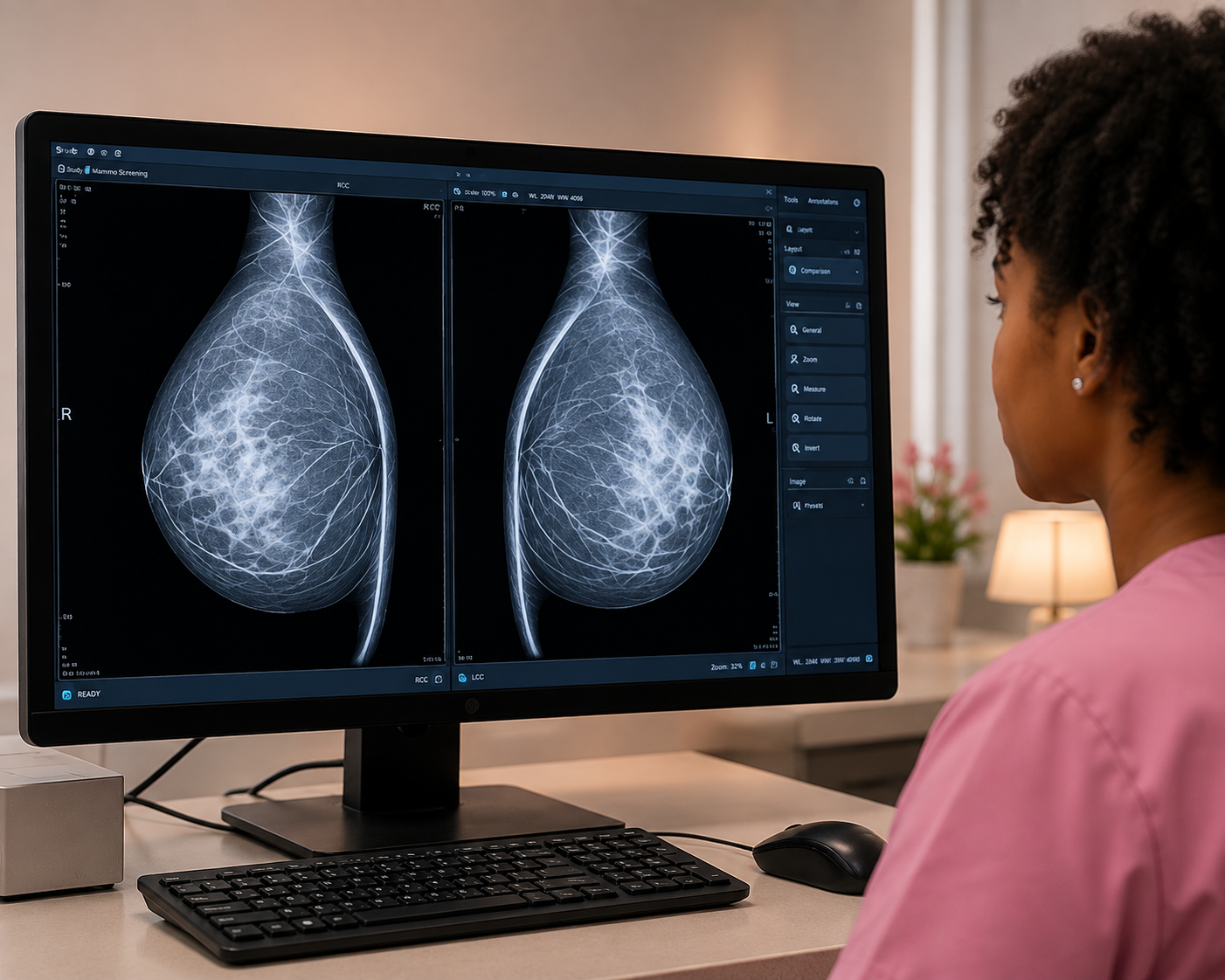
Mammography uses low-dose X-rays to create detailed images of breast tissue. These images help radiologists identify changes such as: Breast lumps, Calcifications Cysts, Abnormal tissue changes, and Early signs of breast cancer.
Digital mammography provides exceptional image quality, enabling accurate diagnosis while using minimal radiation exposure.
Breast Self Examination

Look for: Changes in breast size or shape Skin dimpling or puckering Redness or swelling Changes to the nipple (turning inward or unusual position) Rash or skin irritation Unusual nipple discharge.
Next: Raise both arms overhead and look for the same changes. Place your hands firmly on your hips and tighten your chest muscles while looking for any differences.
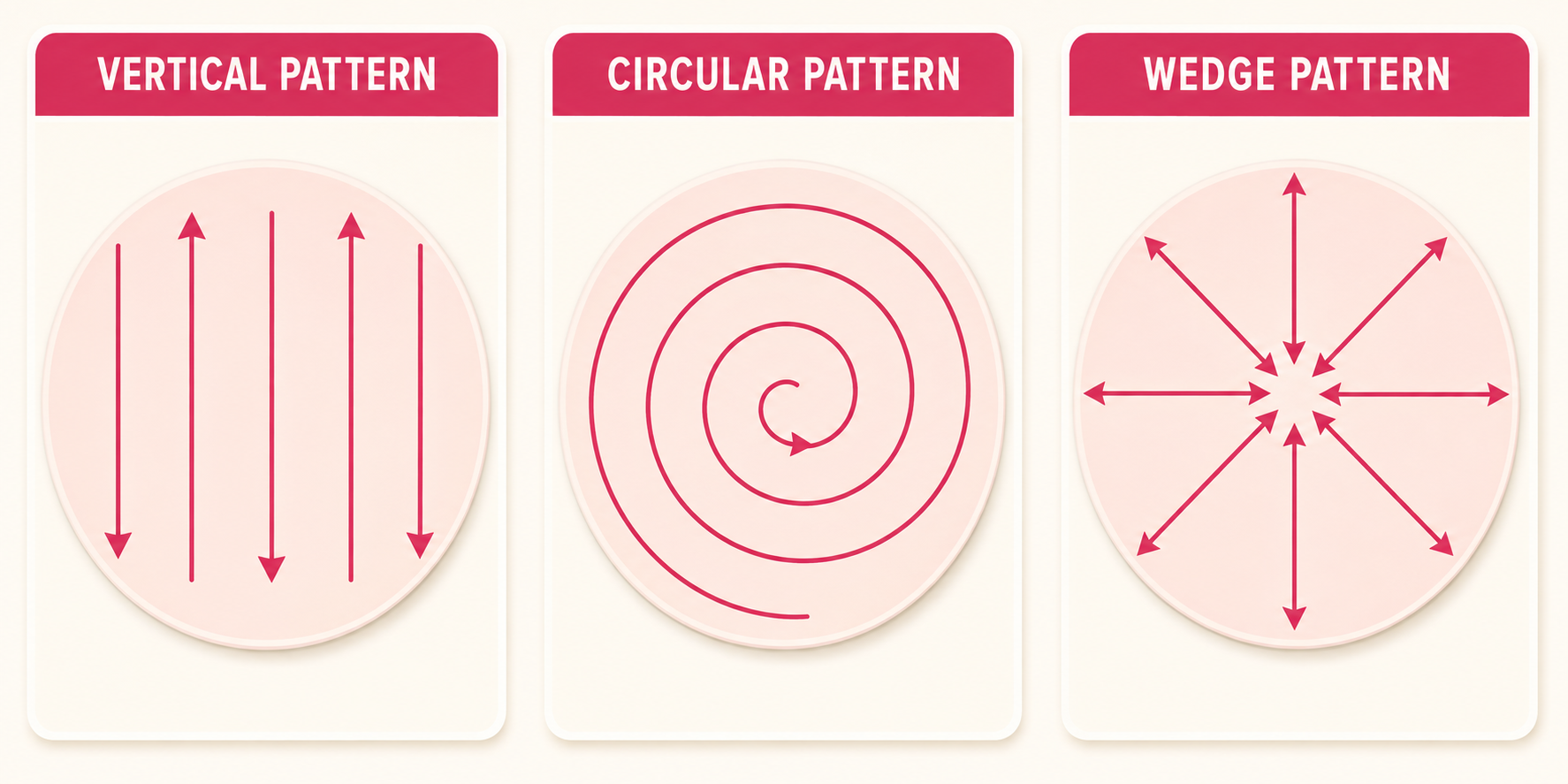
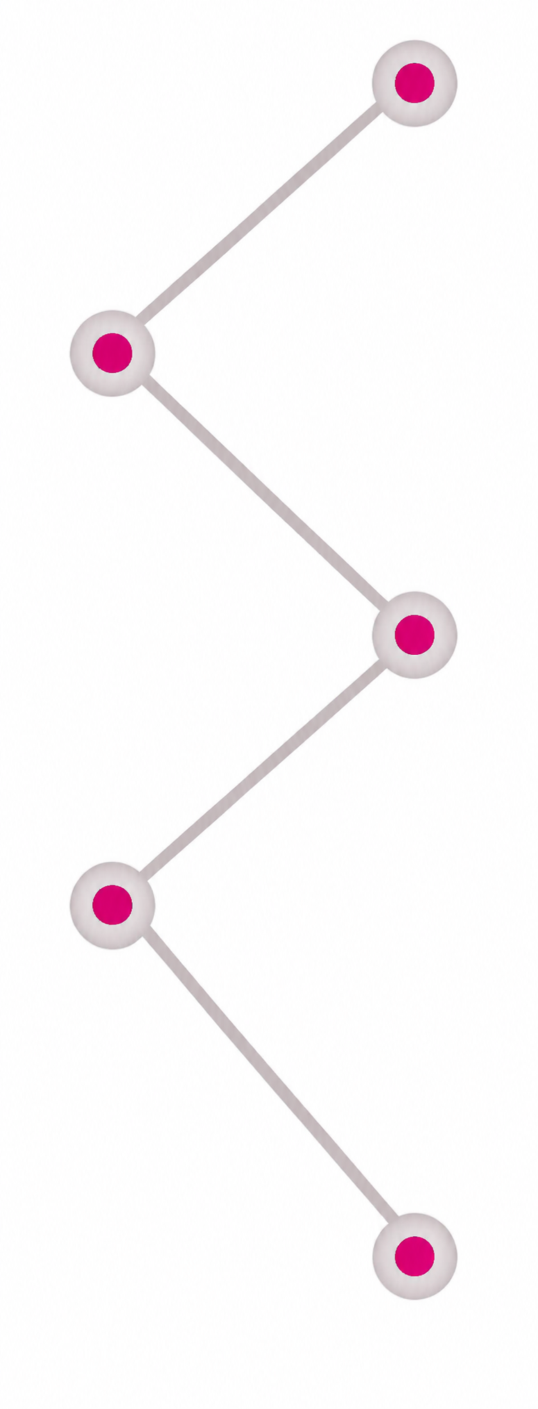
Move your fingers in straight lines: Start at your collarbone. Move down to below the breast. Move slightly inward. Continue up and down until the entire breast has been examined.

Many women find it easier to examine their breasts when the skin is wet and slippery. Repeat the same examination pattern while standing, paying attention to the entire breast and underarm area.

Lie comfortably on your back. Place a pillow under your left shoulder. Put your left arm behind your head. Using the finger pads of your right hand (not your fingertips), examine your left breast.
Apply three levels of pressure: Light pressure – to feel tissue just beneath the skin. Medium pressure – to feel the middle layer of tissue. Firm pressure – to examine the deeper tissue near the chest wall.
Apply Repeat the examination on the right breast using your left hand.

Examine the area under each arm. Feel for: Swelling Lumps Tenderness Enlarged lymph nodes
Frequently Asked Questions
A Mammogram is the name for a specialised X-ray of the breast. Mammograms are carried out by Mammographers, female Radiographers or Assistant Practitioners that have had specialist trained to perform this examination.
During a Mammogram, your breasts are exposed to a small amount of radiation, which is around 0.6millievers (mSv). For comparison, in the UK the annual dose of natural background radiation a person receives is around 2.7 mSv, so the risk to your health is very low.
Mammograms are able to detect changes within your breast, some of which you maybe aware of such as a breast lump. A Mammogram may also detect changes that cannot be felt during a physical examination. This includes small changes within your breast which may require further investigation. The early detection of breast cancer can lead to higher success rates in treatment.
A Mammogram is the best tool for detecting breast cancer for several reasons. One reason is That this test can detect cancer in your breast up to 3 years sooner than you would
notice a lump, it gives doctors or Radiologists a clear image that shows patterns in your breast by monitoring these patterns it enables the doctors to act quickly so that less invasive treatment may be an option as well as giving you more treatment options to begin with. Finding a cancer more rapidly can increase the chance of cancer removal via surgery
There are no special preparations before this procedure. You can eat and drink normally, before and after, however please do not wear deodorant, talcum powder, lotion or perfume on your breasts and underarms as these products will affect the Xray image.
- You may bring a friend or family member with you for support, however please note that they will not be allowed in the examination room with you
- It is advisable to wear clothes that can be easily removed, as you will be asked to undress from the waist upwards.
- If you have breast implants; please inform the Radiographer when you arrive for your appointment.
- If you have a pacemaker; please inform the Radiographer when you arrive for your appointment.
The procedure should last no longer than 15-20 minutes. The Mammographer will call you in to the x- ray room, ask you a few questions about your general health and explain the procedure to you. If you have any additional questions, she will be more than happy to answer them for you.
Once all the questions have been answered and you are happy to proceed with the Mammogram, the Mammographer will ask you to remove all your clothes from the waist up. She will then get you to stand in front of the X-ray machine where she will
position you for your Mammogram.
Each breast, in turn, will be placed on the X-ray machine and firmly compressed by a clear plate. The compression will only last a few seconds and does not cause any harm to the breasts. Compression is needed to keep the breast still and to get the
clearest picture with the lowest amount of radiation possible. Usually two images are taken of each breast but if your breasts are larger than average it may be necessary
to take more than two images of each breast.
Some women find compression slightly uncomfortable and some feel short lived pain. The pain you may feel can depend on several factors:
- Where you are in your menstrual cycle
- Your individual pain threshold
We would encourage you to discuss any pain felt with the Mammographer so that she can try to find ways to make the exam more comfortable for you.
The Mammogram images will be reviewed by a radiologist and a report will be issued to the consul- tant that referred you. Sometimes it is necessary to call patients back for a further Mammogram to examine more closely any areas of change in the breast. This is very common and although may seem alarming at the time, it is normal procedure to help produce the most accurate report.